Keratoconus

What is Keratoconus?
Keratoconus is the leading cause of blindness especially among the youth in East Africa.
A normal cornea (see definition of cornea below) is shaped like a watch glass, but sometimes it starts thinning and bulging; this is known as Keratoconus (Kerato meaning Cornea and Conus like a cone).
In such cases, the cornea cannot perform its main function of focusing light rays entering the eye on to the retina due to its distorted shape.
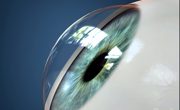
Normal Eye
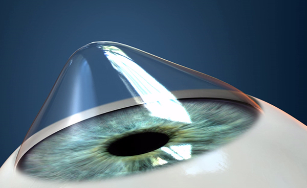
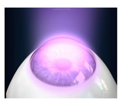
Eye with Keratoconus
Here the middle part of the cornea becomes weak and thin. As a result, it cannot withstand the pressure of the eye causing the cornea to bulge.
The exact cause of Keratoconus is not known, but it is believed that it could be due to a malfunction in the enzymes, which make the stroma (please see cross-section of the cornea) weak. It could also be associated with allergy and there is some evidence that it may be hereditary.
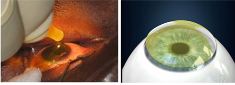
Keratoconus is also linked with over exposure to sunlight, improper contact lens fitting and constant eye rubbing. If there is progressive stretching in the Descemet’s membrane, it leads to rupturing of the membrane and can cause hydrops, where the cornea becomes completely cloudy as shown in the picture below.
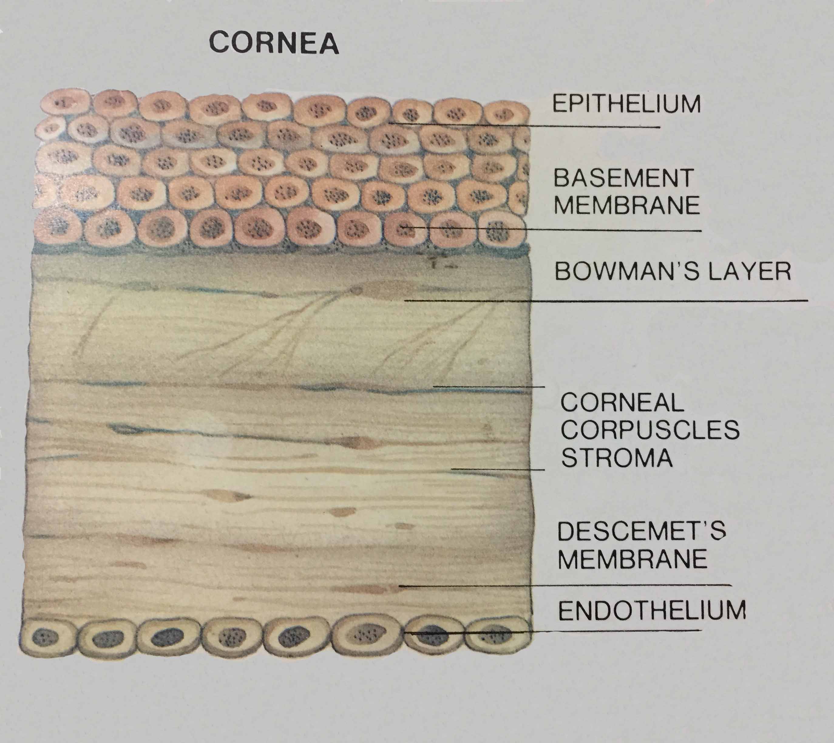
Cross-section of the cornea
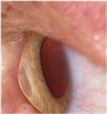
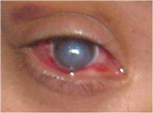
Hydrops – Here the cornea has become competently cloudy
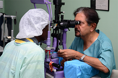
Diagnosis
- Diagnosis of Keratoconus is done through Topography and Tomography, which is a gold standard for Keratoconus but your optician, will suspect Keratoconus if there are following changes:-
- Frequently change in the astigmatism of the eye and despite of giving the correct glasses the vision of the quality is not very good and there is difference in glasses prescription between your two eyes.
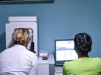
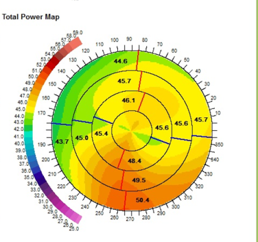
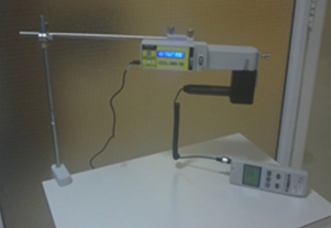
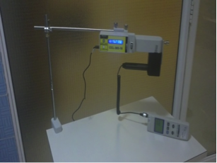
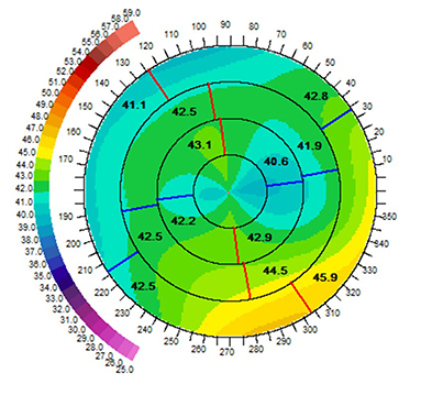
Topography Test
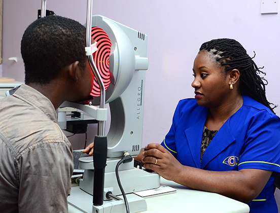
Topography Test
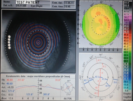
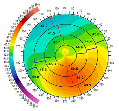
Topography showing Normal cornea
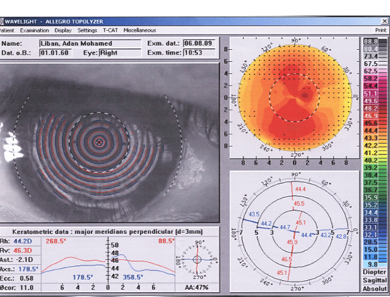
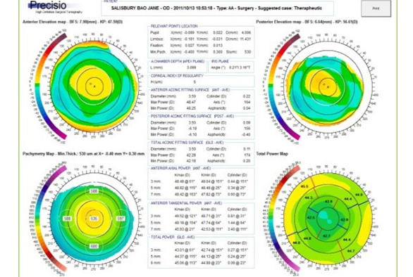
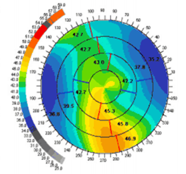
Topography showing Keratoconus
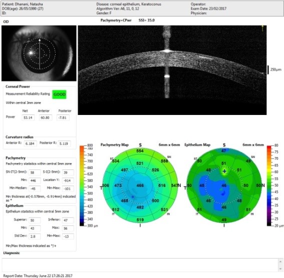
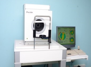
Tomography with the cutting edge Precisio machine
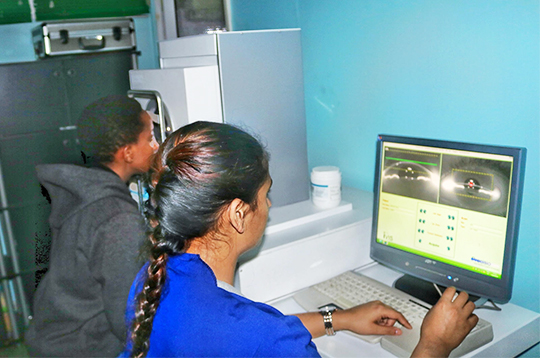
The Topography Machine
Normal Precisio

Precisio Showing Keratoconus
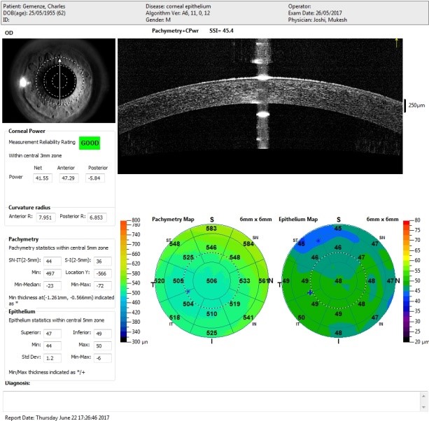
Diagnosis of Keratoconus by Epithelial Mapping
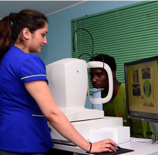
Optical Coherence Tomography test: This test detects early signs of Keratoconus
Normal Tomography test
Tomography showing early signs of Keratoconus
TREATMENT:
At Laser Eye Centre we have the following protocol for treating Keratoconus
- Early to moderate stages we do central corneal regularization followed by Cross-Linking
- Fairly advanced stage only Cross-Linking
- Very advanced stage Corneal Transplant
CENTRAL CORNEAL REGULARIZATION FOLLOWED BY CROSS-LINKING
-
- FIRST STAGE CENTRAL CORNEAL REGULARIZATION
This is done by reshaping the cornea; we reduce the size of the cone by very specialized laser which takes only 34 seconds. - SECOND STAGE COLLAGEN CROSSLINKING:
This is the latest treatment for Keratoconus and was invented by Prof Seiler (Switzerland), and Prof Spoerl (Germany). Cross-Linking has become the standard treatment to treat Keratoconus
- FIRST STAGE CENTRAL CORNEAL REGULARIZATION
CENTRAL CORNEAL REGULARIZATION
- Keratoconus gives rise to irregular irregular Astigmatism and patient’s vision is not improving with glasses and because of atopic conjunctiva contact lenses is a challenge.
- The principle of Corneal Regularization is to reshape the cornea and make the Keratoconus cone as flat as possible and convert the irregular irregular Astigmatism into regular Astigmatism.
- In CCR, the surgeon treats the Keratoconus cone by shifting the cone in the Centre.
- With 1000 Hz frequency, IVIS laser will reshape the top of the cone and the surrounding area.
- This is achieved by performing laser on the side of the cornea which is thicker. Therefore, not damaging the cornea that is already thin.
CENTRAL CORNEAL REGULARIZATION IS DONE IN 34 SECONDS.
CENTRAL CORNEAL REGULARIZATION IS ONLY AVAILABLE AT LASER EYE CENTRE
HOW IS IT DONE?
Step 1:
Checking the mapping of the cornea and diagnosing Keratoconus
Step 2:
Exporting data on to the laser machine
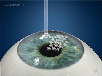
Step 3 :
Central Corneal Regularization in progress
Clinical Results After Central Corneal Regularization
- This is a mini-invasive treatment, which has been proven to strengthen the weak corneal tissues.
- Riboflavin eye drops (vitamin B12) are applied to the patient’s eye for half an hour.
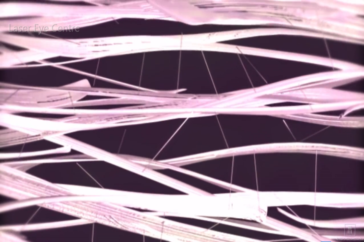
- The Riboflavin is then activated by Ultra Violet light. An interaction between the ultra violet light and the Riboflavin soaked cornea leads to the strengthening and stiffening of the collagen fibers in the cornea.
- This does not allow Keratoconus to progress further. In our clinical practice, cross linking is helpful for 90% of Keratoconus patients but it does not work if the Keratoconus is very advanced.
- The standard length of treatment is 10 minutes.
IF KERATOCONUS IS DIAGNOSED AT AN EARLY STAGE, CROSS-LINKING IS THE TREATMENT OF CHOICE AS IT IS NON–INVASIVE AND RELATIVELY SIMPLE.
- At Laser Eye Center, it is now possible to reduce the cross-linking procedure time to 5 minutes.
- We can do this by incorporating our C-Ten (1000Htz) laser and the CCL-365 platform.
- The overall result is better vision than cross-linking alone in a fraction of the time using cutting edge and safe technology.
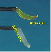
Corneal Strip Before and After Cross linking
After cross linking the cornea has become stiff
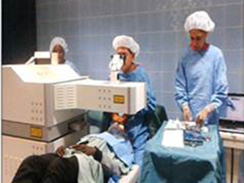
CROSS-LINKING IN PROGRESS
Examining Patient before starting Cross-Linking
Instilling Riboflavin eye drops to the patient’s eyes
Riboflavin Shielding
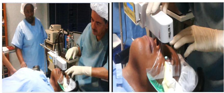
Cross-Linking Treatment in Progress performed by Dr. Mukesh Joshi
CCL 365 10 minute cross linking with 18mW/cm3
UV radiation Light
Side Effects of Cross-linking:-
- If Crosslinking has not been done by an experienced doctor, chances are the crosslinking may not be accurate.
- 90% of crosslinking’s have been successful; however the 10% may require repeating the procedure or may lead to corneal opacity crosslinking.
Spectacles and Contact Lenses:
- With spectacles, vision cannot be corrected due to the irregular shape of the cornea.
- A hard contact lens (rigid and gas permeable) is sometimes a good solution. Here as the lens is made of a hard material it helps to correct the irregular shape of Keratoconus.
- Fitting of these specialised lenses is delicate and time consuming.
- Over time, it can give rise to progressive thinning of the cornea and patients with allergies may have difficulty in contact lens fitting.
If the fitting of contact lens is guided with the help of Topography, the results are excellent.
CCL-365 10 Minute Cross-Linking
CLINICAL RESULTS AFTER CCR AND CXL
Before CCR and CXL
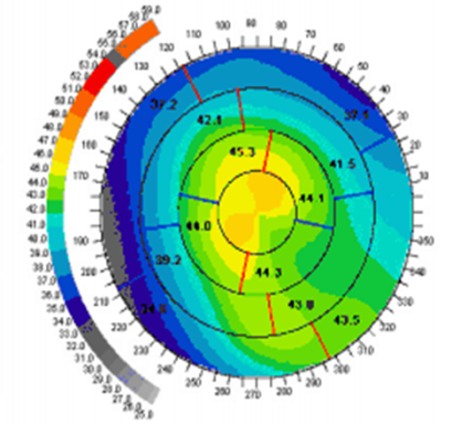
After CCR and CXL
Laser Eye Centre is the first centre in Asia and Africa to offer cross linking treatment in any form and is also the first and only centre to offer the 10 minute Cross-Linking / CCL-365 platform.
- Dr. Joshi was appointed as a global expert on Keratoconus during the 5th International meeting European Society of Cataract and Refractive Surgeons (Germany 2010).
- Dr.Joshi has also been invited by various international societies to share his experience on crosslinking; we at Laser Eye centre were the first to start crosslinking in Africa and Asia.
- An ophthalmologist must undergo training for crosslinking certification so that he/she can do this treatment.
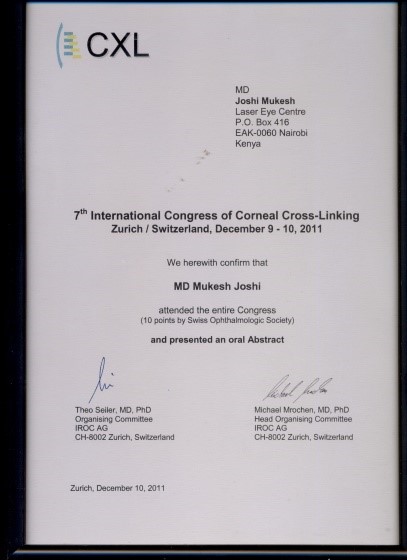
In December 2014, Dr. Joshi was invited as a guest speaker alongside with Prof. Theo Seiler to chair a session at the 10th International Cross-Linking Society meeting.
- Corneal Transplant (also known as Keratoplasty) is usually carried out if the patient’s Keratoconus is at a very advanced stage where contact lenses or cross-linking are not possible.
- The best solution to such cases would be corneal transplant or Keratoplasty (please see the Keratoplasty section of the website).